Background, 망막, 맥락막을 포함하고 있는 11classes Segmentation
- Python 3.6.13
- PyTorch 1.10.2
-
11 classes
- 망막의 경우 총 12개의 layer로 구성
- 막 구조의 ELM(External Limiting Membrane) 제외
- MZ(Myoid Zone), IZ(Interdigitation Zone)의 경우 층이 매우 얇기 때문에 각각 주변층과 하나의 Class로 통합
- Choroid layer추가
-
Dataset
-
각 Volume에 대해서 총 21 Slices이 존재
- Volume : 각 환자의 OD(right eye), OS(left eye)에 대한 OCT Scan Data List
- 황반의 중심을 지나는 11 slice를 기준으로 +-3mm 영역을 관찰하기 위해 5 slice ~ 17 Slice로 총 13개의 B-scan Images이 저장
-
현재 633 Volumes이 존재 >> Total 8229(633*13) Images 존재
-
-
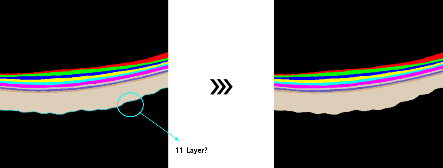
Labeling 과정에서 맥락막과 배경 사이에 새로운 Layer가 잘 못 발생하여 수정
-
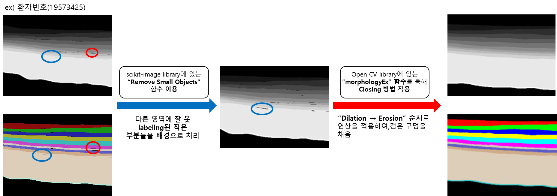
Mask 이미지에 다른 Class로 Labeling 되어있는 부분과 검은색 Hole들 제거
-
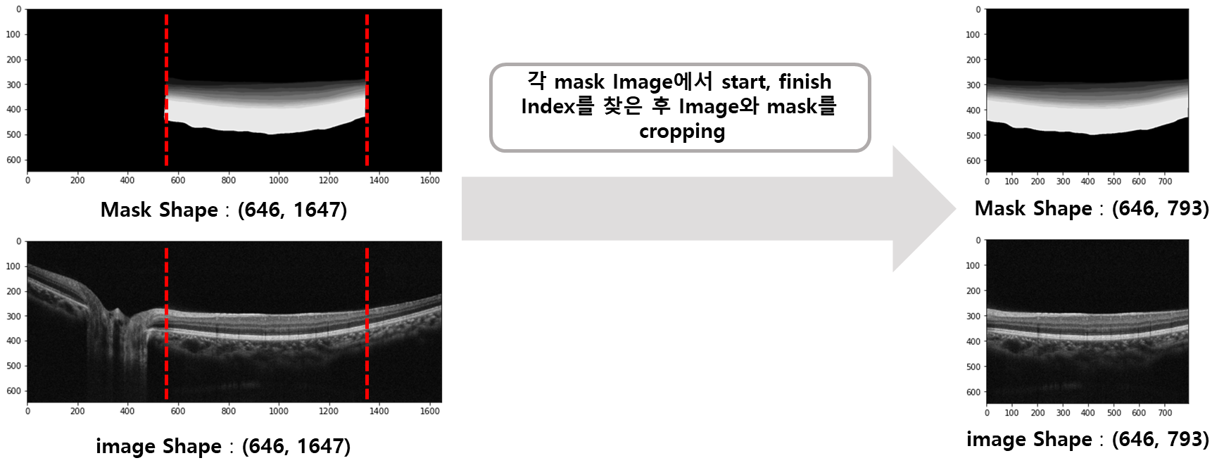
각 Mask Image에서 Start, Finish index를 찾은 후 Image와 Mask를 Cropping
- 각 중증도 별로 Patient를 기준으로 8:1:1 비율로 Split
- Model : Unet (pretrained model)
- Augmentation(Image Augmentation Library, albumnetation 이용)
- Resize, RandomScale, RandomHorizontalFlip, CLAHE
- Epoch : 50
- Optimizer : SGD
- Loss Functioin : CE(Cross Entropy)
- Batch Size : 8
- Learning Rate : 0.001
- 현재 Mask를 기준으로 원본 이미지를 Cropping하였는데, 여기서 기준점 X
- 사전 연구에서는 황반을 중심으로 +-3mm 만큼에 해당하는 pixel 범위의 영역을 사용
- 황반을 찾는 알고리즘 구현
- 원본 이미지에서 edge를 추출
- edge 이미지의 각 열에서 가장 낮은 행 index만 추출
- 이렇게 추출한 line에서 가장 낮은 행 index를 가지고 있는 Pixel의 row, col 찾기
- 황반을 찾는 알고리즘 구현
- 사전 연구에서는 황반을 중심으로 +-3mm 만큼에 해당하는 pixel 범위의 영역을 사용